Factory Seconds
Item: EX10040335
|Model: PHY-HM-4
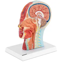
Anatomy Skull - Median Section - Original Size


€89.00
€95.00
Lowest price in the 30 days before the discount: €95.00
Qty
Discount
Per item
3+
4%
€85.44
5+
8%
€81.88
10+
12%
€78.32
Key features
- precise - accurate 3D model of a human skull in original size
- clearly visible - coloured medical model shows the internal structure due to the median section
- universal - ideal as a teaching, decoration or exhibition object
- mobile - can be effortlessly taken to a lecture hall, classroom, etc.
- Accessories - a poster with explanations is included in delivery
Delivery details
Returns and warranty
Downloads
Beautiful piece
The delivery was fast and the product matches the photos very well. I recommend
Product Description
Detailed anatomical skull with muscles and vessels in median section
The 3D model of a skull with a median section PHY-HM-4 from the medical supplies by physa has everything that a realistic replica of the inner and outer structures of the human head requires: All details of the muscles and vessels have been precisely shaped and carefully coloured in their anatomy. It is the ideal model for visualisation and exercises in medical practices, hospitals, schools and universities.
The half skull anatomy model in original size for schools, universities and medical practices
The coloured medical model shows the anatomy of the human skull, on one side with the scalp, superficial muscles, vessels and nerves as well as the auricle and salivary gland. On the other hand, the median section lets you see the interior of the human skull. The brain is shown with all its parts such as thalamus and hypothalamus, pituitary gland, cerebellum, middle and cerebellum as well as the posterior brain, pineal gland and meninges and cerebral fluid in great detail and on an original scale. In addition, the skull anatomy shows the oral and nasal cavity, respiratory and gastrointestinal tract as well as a section of the cervical virtula.
The anatomical model rests on a stable and elegant pedestal that securely holds the skull in place. Since the skull model makes its construction, connections and functions so much easier to understand, it should not be missing from any medicine practice!
As with all high-quality anatomical models from physa, the following also applies here: Important functions as well as diseases can be explained in a comprehensible way with this lifelike 3D model and there is a poster included in delivery. The lifelike visualization of this study object promotes understanding and helps with the learning process, as the three-dimensional model allows for precise analyses of the human skull.
Its particularly realistic shape is made possible by its production from special, lightweight plastic. The particularly robust workmanship of the low-maintenance model highlights the quality of its construction.
Ratings & reviews
Ratings & reviews
4.5
Based on 2 reviews
Sort by :
No comment provided.
Show original review
The delivery was fast and the product matches the photos very well. I recommend
Show original review
Frequently asked questions
How do I clean the model?
To remove dirt from the model, you can easily wipe it with a damp cloth or sponge and a mild detergent.
Is it difficult to build this model?
With this anatomical model you save valuable time! The model is fully assembled, no assembly is necessary!
THE EXPERTS CHOICE
16 YEARS OF EXPERIENCE...
of powering professionals
3 MILLION...
happy and satisfied professional customers