+442037698802
Mo - Fr 8:00 - 14:00
Practice Furnishings
Anatomy Models
Rollator
Item: EX10040257
|Model: PHY-SM-4
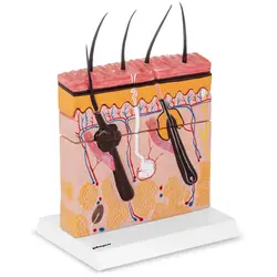
3D Skin Model


€69.00
Qty
Discount
Per item
3+
5%
€65.55
5+
10%
€62.10
10+
15%
€58.65
Back in stock soon
Lowest price guaranteed
Key features
- Medically-accurate anatomical skin model
- Depicts all three skin layers
- Highly-detailed 3D model
- Indispensable in any dermatological practice
- View in cross-section and from above
- Ideal for study of dermatology
Delivery details
Returns and warranty
Downloads
Class
I had a few more questions - they were answered quickly and the delivery arrived safely and well packaged - I would be happy to do so again
Technical details
Material
Plastic
Colour
Coloured
Dimensions (LxWxH)
16 x 22.3 x 31 cm
Weight
1.31 kg
Shipping dimensions (LxWxH)
28 x 18 x 22 cm
Shipping weight
1.9 kg
Delivery package
- 3D Skin Model PHY-SM-4
- Sponge
- Plastic bag
Product Description
3D Skin Model
The three-dimensional skin model PHY-SM-4 from Physa depicts a highly-magnified representative cross-section of the human skin structure. All three skin layers are shown in all their details, including hair follicles, sweat glands, fatty tissue and lymphatic vessels. The medical model is perfectly suited for use as a medical model for visualising and teaching.
This anatomical model offers a magnified view of the human skin and its structure. All details are precisely reproduced and can be accurately analysed and examined using this model. It is ideal for hospitals, doctor's offices, schools and universities. The model's three-dimensionality allows you to discover two views of the human skin structure: in cross-section and from above. The cross-section replicates the structure of the three skin layers, which lie on top of each other. These skin layers can each be individually removed and analysed from above. The skin model provides a variety of possibilities for examination.
All three skin layers are replicated in detail in this three-dimensional anatomical skin model, allowing you to consider the structure of the epidermis, dermis and subcutaneous tissue in detail. The magnified skin section represented by this model depicts both the structure of the individual skin layers as well as the interior structures, including the structure of a hair and a sebaceous gland, in detail.
The three-dimensional skin model is indispensable in any dermatological practice. With this model, doctors can vividly explain important functions of the skin as well as various dysfunctions and diseases to their patients. The skin model is also ideal for use as a dermatological study aid. Students can use this 3D model to examine the structure of all parts of the skin, as well as analyse all relationships and functions of the various glands, vessels, hair follicles and tissues.
The 3D skin model comes with a sturdy plastic stand which keeps the model stable. The stand also allows you to attractively display and securely store the 3D model. The stand keeps the model steady and allows it to be used even during hectic school or hospital operation.
The study aid is quick and easy to assemble: Just lay the individual skin layers on top of each other and position them on the included stand. You don't need any tools or even much time. All the elements of this skin model are depicted in different colours, making it easy to differentiate between the individual parts of the skin. The distinctive colouring also underlines the detailed 3D model's high degree of realism.
Ratings & reviews
Ratings & reviews
5
Based on 1 review
Sort by :
I had a few more questions - they were answered quickly and the delivery arrived safely and well packaged - I would be happy to do so again
Show original review
Frequently asked questions
Is the skin model difficult to put together?
Thanks to its user-friendly design, the skin model is quick and easy to assemble.
How do I clean the skin model?
To clean the skin model, simply wipe it down with a moist cloth or sponge and some cleaning agent.
THE EXPERTS CHOICE
17 YEARS OF EXPERIENCE...
of powering professionals
3 MILLION...
happy and satisfied professional customers